Basal Cell Carcinoma (BCC, rodent ulcer)
Read - The value of Mohs surgery for nmsc in J cutan aesthet surg 2012 5(1) 1-2
Read - our Facial basal cell carcinoma publication in the BMJ (free)
BCC patient information leaflet
BCC guidelines November 2021
Basal cell carcinoma (BCC) is a slow growing locally invasive skin cancer. There were estimated to be 410,716 new cases in 2015 and the incidence is increasing at 2-5%.
BCC is the commonest cancer of fair skinned populations (ref. 1)
One of the reasons is sun exposure. Thinning of the ozone and increased travel to sunny climates may be contributing factors. BCC represents approximately 80% of all skin cancers. Several factors are responsible for BCC causation including cumulative sun exposure, fair skin type, sunbed usage, smoking, sunburn and immunosuppressive treatment for an organ transplant.
BCC is generally slow growing and slowly invades local skin and surrounding structures. Most are relatively benign and are more of a nuisance than a threat to one’s health. Spread to other parts of the body (metastasis) is very rare. BCC’s occur mainly on sun-exposed sites of fair-skinned individuals, the most common location being the head and neck. Sometimes large or neglected BCCs are capable of extensive local destruction and invasion ( “rodent” ulcer) – gnawing their way through skin, muscle, bone. Fortunately BCC rarely kills people or harms their health as most as small and easily treated by a number of methods. However, as most occur on the face, having facial surgery with subsequent scarring is not to be taken lightly. In some cases, skin cancer removal can be psychologically distressing and can result in physical disfigurement.
CLINICAL
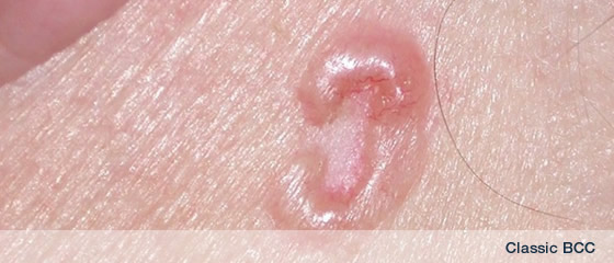
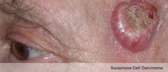
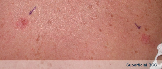
BCC has a variety of clinical presentations; most are small (approximately 1cm in diameter), well-defined, red or “pearly” or flesh-toned lumps. There may be a central ulcer encircled by a rolled edge with surrounding telangiectasia (fine blood vessels). The BCC can mimic an ulcer or be a raised lump. The BCC may bleed followed by crust formation which separates to reveal bleeding and then crusts over again. They are described by some as a “non-healing” sore. Following a history and examination, a biopsy will establish the diagnosis. The four major types of BCC are nodular, superficial, morpheic [infiltrative] and pigmented. (ref.1).
The usual symptoms of a BCC are a slowly growing lump or patch or plaque. This often is like a non-healing sore spot. Often the area will bleed, followed by scab formation and it will appear to be healing, but then the scab is dislodged by touch or after showering or towelling and the lesion then bleeds. The lump may appear pearly, translucent, pinkish or red. The edges of the lump may be rolled and the centre may be depressed, sunken or ulcerated with scab formation. If it is a patch then it may appear like a patch of eczema or psoriasis.
If you notice a non-healing sore spot or a lump that grows gradually or it has a pearly appearance then consult your doctor. If you have a patch of what looks like eczema but it doesn’t improve with creams or ointments used in eczema management then see your doctor.
Reference:
1. Ceilly RI, Del Rosso JQ. Current modalities and new advances in the treatment of basal cell carcinoma. Int J Dermatol 2006; 45: 489-98
Standard Treatment of BCC
| 0. |
No treatment is an option but no treatment of BCC runs the risk of significant progression of the tumour in at least 25% over 2-5 years |
| 1. |
Local anaesthetic surgery with predetermined margins – also known as wide local excision (WLE) |
| 2. |
Mohs micrographic surgery |
| 3. |
Cryotherapy |
| 4. |
Imiquimod |
| 5. |
Curettage and cautery |
| 6. |
Photodynamic therapy |
| 7. |
Radiotherapy |
Surgical Excision = Wide local excision = WLE
This is highly effective for BCC. For well defined BCC <2cm, excision margins of 4mm will result in a 95% clearance rate. BCC >2 cm may require wider margins to effect clearance. Recurrent BCC require wider excisional surgery or Mohs micrographic surgery.
Excision of a BCC under local anaesthetic with 4mm margins or more is a standard treatment. The procedure takes 30-60 minutes depending on the complexity of the cancer but is usually straightforward. Stitches are usually in place for 5-7 days.
Pre-op
 |
|
 |
|
 |
|
|
| BCC |
|
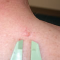
Classic nodular bcc with telangiectases |
|
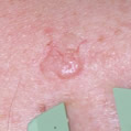
nBCC ready for excsion with 4mm margin |
|
|
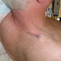
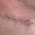
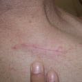
Post-op
 |
|
 |
|
 |
|
|
| Immediately post-op |
|
Close up post-op |
|
Scar 2-3 months post-op |
|
|
Mohs micrographic surgery
Mohs micrographic surgery is the gold standard but is not necessary for the vast majority of BCC. It is a specialised form of surgery usually reserved for cancers that have recurred or where tissue sparing is advantageous or for high risk BCC or where re-excision of an incompletely excised BCC would be a major problem – eg eyelid. It is the treatment of choice for certain rarer cancers such as dermatofibrosarcoma protruberans.
Mohs micrographic surgery (MMS) is a much more precise method of excising a tumour than excisional surgery with a pre-determined margin = wide local excision = WLE. The tumour is excised, divided and stained. Examination of horizontal sections examines 100% of the excised tumour margin. However MMS is time consuming, costly and requires specialist expertise. (see link to SKCIN Trustee Richard Clifford having Mohs micrographic surgery of his cheek BCC)
Curettage and cautery
This technique is best reserved for small, well-defined BCC on low risk sites.
Cryotherapy
This is a quick, easy, inexpensive and effective treatment which can result in excellent cosmesis. In reality cryotherapy is not used much for skin cancers except in selected circumstances. Depending on the patient, lesion and operator experience, high cure rates can be gained.
Radiotherapy
A useful treatment for BCC or patients not suitable for surgery. Radiotherapy is an excellent option for certain skin cancers. In skilled hands, it offers high cure rates with sometimes excellent outcomes
Photodynamic therapy (PDT)
PDT involves the application of a photosensitising prodrug to the skin cancer. After soaking into the cancer for 4-6 hours, this gets converted to a photosensitser. Activated by an appropriate light source produces a local reaction killing off the cancer. Currently PDT for BCC is reserved for BCC unsuitable for treatment by other methods or where the cosmetic outcome may be considered more favourable to surgery. Good cure rates with excellent cosmetic results can be gained for selected BCC.
Imiquimod
(Aldara) is a cream that is licensed for the treatment of superficial BCC. This cream is useful for low-risk BCC in non-critical sites. Applied 5x/week for 6 weeks, it evokes an inflammatory reaction. The inflammatory response destroys the lesion. Considerable soreness can occur. Assessment of response takes place after a further 12 weeks. For selected BCC, this is a good option with good cure rates but long term follow up data is sparse.
Pembrolizumab shows promise in aBCC
|