Mohs Micrographic Surgery
What is Mohs surgery?
Read - The value of Mohs surgery
This is the Gold standard treatment for facial skin cancer
| 1. |
Ensures complete cancer removal |
| 2. |
Preserves healthy tissue |
| 3. |
Aesthetics are important on the face so offers better options at closure of defect |
Read our Facial basal cell carcinoma publication in the BMJ (free)
Watch a video from OHSU on Mohs surgery
Watch a video on Mohs surgery
Learn more about Mohs surgery
See Mohs surgery in pictures
The aim of surgery is to completely remove skin cancer.
Mohs micrographic surgery is named after Dr. Fred Mohs who described it in 1941 and is a specialized procedure used to remove skin cancer. The procedure involves microscopic analysis and a map (graph = micrographic) of a tumour to ensure that all the skin cancer is completely removed. This gives the best chance of total eradication of the cancer and ultimately a cure from that skin cancer.
This is not a routine procedure but reserved for those tumours designated as being difficult, more aggressive, large, unusual, recurrent, previously incompletely removed or located at cosmetically sensitive or anatomically important sites.
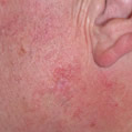
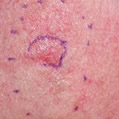
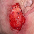
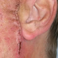
Examples of Mohs Micrographic Surgery:
Pre-op
 |
|
 |
|
 |
|
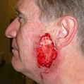 |
| BCC on cheek |
|
BCC extent marked out |
|
Defect after 4 stages of Mohs surgery |
|
Defect |
Post-op
 |
|
 |
|
|
|
|
| Closure |
|
Post-op |
|
|
|
|
For most skin cancers, simply cutting out them out or curetting (scraping) them may be sufficient for complete removal depending on the site, size and sub-type of tumour.
Mohs surgery is a much more precise and accurate way of tumour removal. It offers immediate and complete histological examination of the surgical margins. No other treatment modality approaches the consistently high cures rates for non melanoma skin cancer (NMSC, such as BCC and SCC) offered by Mohs surgery. (ref.1)
Additionally, Mohs micrographic surgery spares normal tissue. Thus, it offers the highest cure rates and aims to create the smallest surgical wound which is advantageous because this offers the surgeon sometimes simpler or more reconstructive options than if a larger wound had been created.
5-year cure rate for primary BCC (ref. 2)
| Mohs |
99% |
| Excisional surgery |
89.9% |
| Cryotherapy |
92.5% |
| Curettage and cautery |
92.3% |
| Radiotherapy |
91.3% |
Mohs micrographic surgery is cost effective (ref.3)
Reference
1. Otley CC. Non-melanoma skin cancer. Past Present and future. Curr Probl Dermatol March/ April 2001; 109-113.
2. Rowe DE, Carroll RJ, Day CL Jr. Long-term recurrence rates in previously untreated (primary) basal cell carcinoma: implications for patient follow-up. J Dermatol Surg Oncol 1989; 15: 315–328.
3. Cook J, Zitelli JA. Mohs micrographic surgery: a cost analysis. J Am Acad Dermatol 1998; 39: 698-703.
Watch a video from OHSU on Mohs surgery
Watch a video on Mohs surgery
Learn more about Mohs surgery
See Mohs surgery in picture
|