Squamous Cell Carcinoma (SCC)
SCC patient information leaflet
SCC 2020 guidelines published March 2021 BJD
Read our Scc article
Panthagani A, Varma S. A practical guide to squamous cell carcinoma and other non-melanoma skin cancers. Dermatology in Practice 2018; 24(2): 32-38.
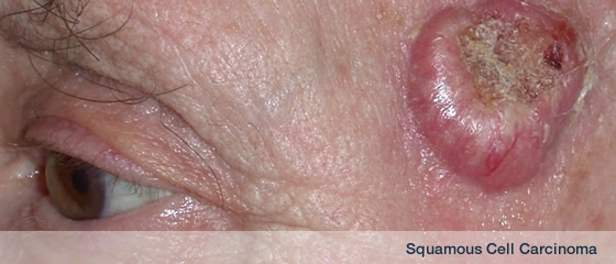
What is a squamous cell carcinoma?
This is the second most common type of skin cancer and is most commonly found in older people, but the popularity of sunny holidays and recreational use of sunbeds means that we are seeing them in younger age groups. SCC is a cancer of the keratinocytes of the skin.
| • |
The incidence is continuing to increase. |
| • |
At Kings College in the United Kingdom, a 10-fold increased incidence rate was seen in SCCs from 1970 to 1992. (ref.1) |
| • |
In 2016 a report from Norwich noted that patient numbers with SCC had increased 2.8 fold (Goon KC, Greenberg DC, Igali L, Levell NJ. Squamous cell carcinoma of the skin has more than doubled over the last decade in the U.K. Acta Derm Venereol 2016; 96: 820-21. |
The most likely sites are the areas of skin often exposed to the sun, typically the face, ears, lips, mouth and hands. The appearance varies but is usually a scaly lump, nodule, ulcer or non-healing sore. They often start as small hard white or skin-coloured lumps in the skin that grow at a variable rate. Squamous cell carcinomas have the ability to spread to other parts of the body, but do not often do so. If left untreated, the tumour will increase in size and damage the surrounding skin. It may then spread to local lymph nodes or around the body and ultimately kill an individual.
Reference
1. Hughes JR, Higgins EM, Smith J, Du Vivier AW. Increase in non-melanoma skin cancer: the King’s College Hospital experience (1970-92). Clin Exp Dermatol 1995;20:304-7).
What causes a squamous cell carcinoma?
The main cause is exposure to sunlight over many years. Occasionally, a squamous cell carcinoma may be caused by exposure to certain substances used in industry, such as tar. They can sometimes develop in chronic ulcers or in patients who are on drugs that suppress the immune system.
Risk factors for SCC
| • |
UVR – excessive sun exposure |
| • |
PUVA - photochemotherapy |
| • |
OTR immunosuppression |
| • |
Organ Transplantation recipient - OTR are more at risk of developing skin cancer compared to other patients |
| • |
Chronic ulcer |
| • |
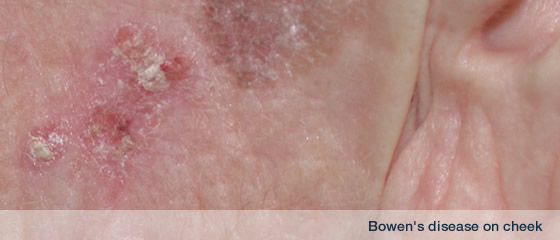
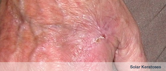
Precancerous lesions such as Actinic keratoses or Bowens disease |
Treatment of Squamous cell carcinoma
| 1. |
WLE = wide local excision |
| 2. |
Mohs micrographic surgery |
| 3. |
Radiotherapy |
In the majority of cases, the SCC can be removed under local anaesthetic on one visit
Surgical Excision
For well defined tumours, <2cm in diameter, excision with at least 4mm margin completely removes the tumour if 95% of cases.
Larger tumours, thicker tumours, those on high risk sites or those with poor differentiation should be excised with wider margins
Radiotherapy may be appropriate for
| • |
selected individuals |
| • |
certain lesions |
| • |
as an alternative to surgery |
SCC has the ability to spread around the body (metastasize) and therefore kill.
Certain factors predict the ability of an SCC to metastasise
| • |
Size >2cm |
| • |
Site - ear, lip |
| • |
host immunosuppression |
| • |
Perineural invasion |
| • |
Differentiation - poorly > moderately > well Depth -thicker >thinner |
| • |
Aetiology- non-sun exposed sites, areas of chronic inflammation |
| • |
mucosal SCC > cutaneous SCC |
Overall 5 year survival - cutaneous SCC - 75%-90%.
For metastatic disease - 25%.
Note !
Scc with >4mm/month growth rate distinguishes a subset of high risk SCC with higher risk of lymph node LN spread and a shorter time to LN metastasis. High growth rate was inversely correlated with tumour age.
Authors concluded that GR is more important than age or tumour size. [Canueto J et al. rapid growth rate is associated with poor prognosis in cutaneous squamous cell carcinoma. Clin Exp Dermatol 2018; 43: 876-82]
Two independent tumour staging systems for SCC exist that use high risk features to predict behaviour
AJCC 8
AJCC 8 now revises staging by taking into account several prognostic factors
T1 <2cm
T2 >2 cm <4
T3 >4, bone erosion, PNI, deep invasion Depth> 6mm Or beyond s/c fat , PNI - nerve deeper than dermis or >0.1mm
T4 cortical bone erosion or skull base involved
High risk >=2 cm
PNI - nerve deeper than dermis or >0.1mm
Depth> 6mm
Or beyond s/c fat
BWH - Brigham and Women's hospital
Four high risk features
- Diameter >=2 cm
- Poorly diff
- PNI >=0.1mm
- Invasion beyond s/c fat
T1 0 high risk factors
T2a 1 high risk factors
T2b 2-3 high risk factors
T3 >=4 high risk factors or bone inv
SCC2020 guidelines were published by the BAD in the March 2021 BJD
Lymph node exam
cSCC depth
<2mm - little metastatic potential
2.1-6. - 4%
>6. 16%
Tumours involving fat =up to 46%
Parotid is the site of approx 70% metastatic cSCC of head and neck
Newlands et al. J laryngol oncol 2016 130 s125-132 |