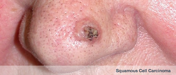
Mohs surgery has the highest cure rates for skin cancer and keeps defects as small as possible by removing the smallest amount of normal skin possible.
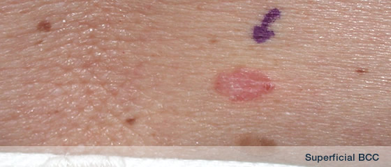
It is also used for poorly defined skin cancers where microscopic analysis is vital to make sure all the “roots” (subclinical invisible cancer) have been removed.
It is a specialised technique which is labour intensive and requires at least 1 year of training. I undertook my training with Dr. Neil Swanson and Dr. Ken Lee in Portland, Orgeon, USA in 2000-2001.
 |
|
 |
|
 |
|
 |
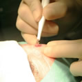
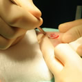
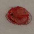
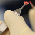
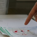
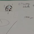
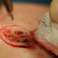
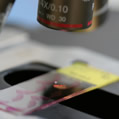
| Ill defined bcc being debulked |
|
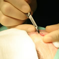
The 1st layer of Mohs surgeryt |
|
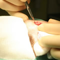
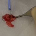
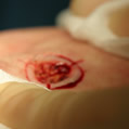
Blade angled at 45°, narrow margin around debulk |
|
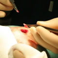
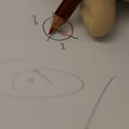
Hatch marks applied |
| |
|
|
|
|
|
|
 |
|
 |
|
 |
|
 |
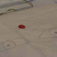
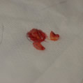
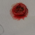
| Tissue removed |
|
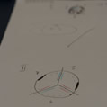
Placed on map. |
|
Close up of removed tissue |
|
10. tissue being divided into sections |
| |
|
|
|
|
|
|
 |
|
 |
|
 |
|
 |
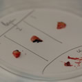
| Excised specimen divided into 3 smaller specimens |
|
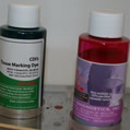
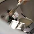
Dyes to ink margins |
|
Specimen edges being inked |
|
Precise map to correlate with dyed specimens |
| |
|
|
|
|
|
|
 |
|
 |
|
 |
|
 |
| Inked specimens & debulked tumour |
|
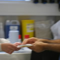
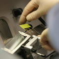
Handed to pathology technicians |
|
Discussion with technicians regarding specimes |
|
Specimen fozen, mounted on chuck ready for making into slides for microscopic analysis |
| |
|
|
|
|
|
|
 |
|
 |
|
 |
|
 |
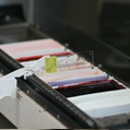
| Technicians cut specimens |
|
Slides being stained |
|
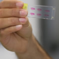
Slides viewed for quaility |
|
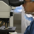
Slides viewed microscopically |
| |
|
|
|
|
|
|
 |
|
 |
|
 |
|
 |
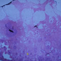
| Cancer seen |
|
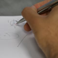
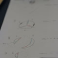
Positive areas marked out on map |
|
Cancer seen in the 1st layer marked on map - this guides accurate removal on layer 2 |
|
Going back to the patient to accurately take a wider and deeper margin |
| |
|
|
|
|
|
|
 |
|
 |
|
 |
|
 |
| 2nd layer |
|
New precise hatch marks |
|
Process repeated, sections, slides, microscopic anlaysis |
|
2nd layer shows cancer at edges so a 3rd layer will be taken just at the positive edges |
| |
|
|
|
|
|
|
 |
|
|
|
|
|
|
| A 3rd and 4th layer were needed to clear this small but ill-defined BCC |
|
|
|
|
|
|