Recognising Skin Cancer
Read our Facial basal cell carcinoma publication in the BMJ (free)
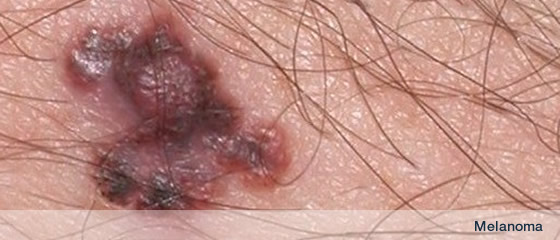
Skin cancer can be thought of as Malignant Melanoma and Non-Melanoma skin cancer.
NMSC comprises basal cell carcinoma and squamous cell carcinoma. Bowen’s disease and actinic (solar) keratoses are pre-cancers that can turn into squamous cell carcinoma.
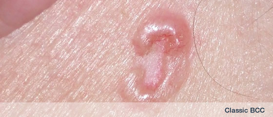
Recognising Basal cell carcinoma
CLINICAL
The typical patient has fair hair, light coloured eyes and fair skin. These individuals may have spent a considerable part of their lives outdoors (living overseas, occupation or hobbies). BCC has a variety of clinical presentations; most are small (approximately 1cm in diameter), well-defined, erythematous, “pearly” or flesh-toned papulonodules or plaques. There may be a central ulcer encircled by a rolled edge with surrounding telangiectasia. The BCC can mimic an ulcer or be exophytic. The BCC may bleed followed by crust formation which separates to reveal bleeding and then crusts over again. They are described by some patients as a “non-healing” sore. They can become fibrotic and resemble a scar. The varying appearances have led to BCC being described as superficial, nodular, cystic, nodulo-ulcerated, morphoeic (sclerosing), keratotic or pigmented. Because most are slow growing and cause few symptoms, patients may not present for several years and may lead to the BCC being neglected. Large or neglected BCC can behave like a “rodent” ulcer gnawing their way through skin, muscle, bone & meninges. Following a history and examination, a biopsy will establish the diagnosis.
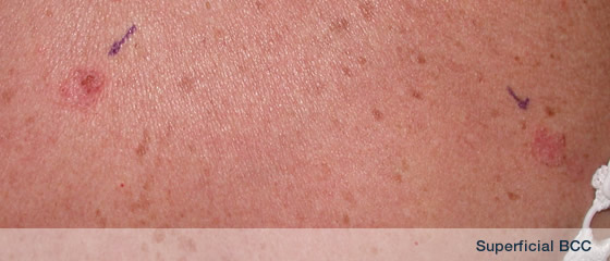
Superficial BCC
This presents as an erythematous patch or plaque, most commonly on the trunk with fine scale and visible telangiectasis. The BCC may be slightly ulcerated, show central fibrosis and have an ill defined geographic border. Differential diagnosis includes nummular eczema, psoriasis, Bowen’s disease, tinea corporis or mycosis fungoides.
Nodular BCC
This presents as round, spherical, oval or dome-shaped papule or nodule with a pearly translucent appearance. Typically soft to firm in consistency, it exhibits slow growth and may ulcerate centrally (“rodent ulcer”) or appear cystic. Differential diagnosis includes an intradermal naevus or sebaceous hyperplasia.
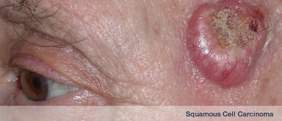
Recognising Squamous Cell Carcinoma
Individuals at greatest risk are fair skinned with excess sun exposure. Sun-exposed sites such as the head and neck, dorsae of the hands and legs are most commonly affected. SCC can arise de novo or from sites of chronic skin inflammation such discoid lupus erythematosus, old burn scars, sinus tracts, chronic leg ulcers and lupus vulgaris (cutaneous tuberculosis) and these SCC are subject to higher rates of metastases. SCC can develop from precursor lesions such as actinic keratoses (AK) or Bowen’s disease (BD) and have the potential for invasion and metastases. Immunosuppression significantly increases the risk for SCC.
CLINICAL
Patients will present with a firm, flesh-toned, endophytic or exophytic indurated papule or nodule or a “non-healing lump” which is sore, painful, oozes, bleeds or is enlarging rapidly usually on a sun-exposed site. The lesion may be smooth, have a scaly surface and be ulcerated, crusted or hyperkeratotic. If infected, SCC can be malodorous or fungating. Following history and examination, a biopsy should include the base so that invasiveness and thickness can be established.
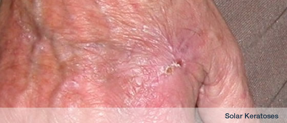
Recognising Actinic Keratoses
Actinic keratoses (syn. solar keratoses, AK) are pre-malignant skin lesions with the potential to develop into SCC. AK are the result of long-term sun overexposure. They are extremely common, especially on the sun-exposed skin of fair-skinned Caucasians. More than 80% occur on the back of the hands, forearms and on the head and neck. Advancing age, male sex, outdoor occupation or hobbies are all additional risk factors.
CLINICAL
Usually AK appear as small, single or multiple scaly erythematous papules smaller than 1cm in diameter. They may be flesh-toned, pink or brown and typically present on sun-exposed sites. Patients may complain of them as being rough, causing soreness, irritation, discomfort or pain or just posing a cosmetic nuisance.
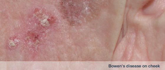
Recognising Bowen’s Disease
Bowen’s disease is an intraepidermal carcinoma in-situ. This is a precancer and in-situ refers to the fact that the disease has not penetrated the basement membrane. Once this occurs, the lesion is best described as a squamous cell carcinoma.
CLINICAL
Clinically Bowen’s disease (BD) presents as an asymptomatic slow growing, usually solitary, sharply demarcated, scaly erythematous patch or plaque. Differential diagnosis includes psoriasis, nummular (discoid) eczema, lichen simplex chronicus, actinic keratoses, superficial BCC or SCC. The surface may be flat, scaly, eroded, velvety or verrucous. Common sites are the lower limbs and head and neck. Women are affected more than men in the UK. Definitive diagnosis is established by a biopsy, usually a punch biopsy. Biopsy through the clinically thickest part of the lesion to rule out invasive SCC.
|