Malignant melanoma
As Lead Clinician for skin cancer in Nottingham [Jan 2012 - Current] I have been dealing with increasing cases of melanoma over the past few years.
A melanoma usually arises from a new mole or an old mole that changes. If a melanoma is caught early and cut out, this can sometimes effectively be a "cure" . If you have moles that you are worried about and want to have an assessment click Appointments.
If you've been diagnosed with a melanoma then click Melanoma Diagnosed.
Cutaneous malignant melanoma (MM) is a cancer of the pigment cells of the skin (melanocytes). The number of new melanomas is continuing to increase in the UK.
There are approximately 16,200 new cases of melanoma in the UK each year with approximately 2000 deaths.
http://www.cancerresearchuk.org/about-cancer/type/melanoma/
There have been enormous advances in melanoma diagnosis, treatment and care over the past 5 years.
Check out
Melanoma: assessment and management NICE
AJCC staging 8
Cancerresearchuk.org for a superb resource and up to date information
Risk Factors
| • |
Fair skin with an inability to tan (skin types I and II) |
| • |
UV radiation = excessive sun exposure |
| • |
Sunburn |
| • |
Burning or high sun exposure at a young age |
| • |
Sunbeds |
| • |
Atypical or irregular moles |
| • |
Lots of moles >100 in number |
| • |
Red hair |
| • |
Numerous freckles |
| • |
Family history of melanoma |
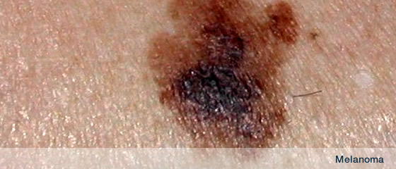
Remember the ABCD(E) of Melanoma
| A |
asymmetry |
| B |
irregular border |
| C |
irregular colour |
| D |
diameter over 7mm |
| (E) |
elevation |
Always get a pigmented lesion assessed if there has been rapid or a recent change in size, shape or colour, inflammation, oozing or bleeding or a change in sensation or if the mole is new and there is rapid change
There are 4 main types of melanoma:
| 1. |
Lentigo maligna (LM) |
| 2. |
Superficial spreading (SSMM) |
| 3. |
Nodular |
| 4. |
Acral lentiginous (ALMM) |
Lentigo maligna (LM) and Lentigo maligna melanoma (LMM)
Lentigo maligna = LM = Hutchinson’s melanotic freckle can be considered to be a precancerous freckle
Lentigo maligna is the premalignant, precancerous or in-situ phase of malignant melanoma.
LM demonstrates a long growth phase, staying precancerous for years before progressing through peripheral extension until a raised central nodule of full blown cancerous malignant melanoma arises = Lentigo maligna melanoma (LMM).
LM occurs most commonly on the face of the elderly.
Superficial spreading malignant melanoma (SSMM)
This is the commonest melanoma in fair skinned individuals.
Any site is possible but malignant melanoma commonly affects sun exposed sites such as the back, chest, arms and legs
| • |
Men are as likely as women to acquire melanomas on their lower legs. |
| • |
Women are as likely as men to acquire melanoma on their upper backs |
Nodular melanoma
This type of malignant melanoma is a nodule and can arise anywhere on the body. These tend to grow more rapidly than SSMM and can present late when they start to catch on clothing or ulcerate and bleed.
Acral lentiginous melanoma (ALMM)
These occur on acral sites (limbs or extremities) such as the palms, soles and under the nail.
EXAMINATION
| Melanomas show |
| Asymmetry in shape or colour distribution |
| Irregular Borders |
| Different Colours |
| Diameter >7mm |
| (E)nlargement or (E)levation |
Prognosis / survival in malignant melanoma / Breslow thickness
The long term outcome from malignant melanoma depends on its thickness called the Breslow thickness. This is measured microscopically when a pigmented lesion is excised.
It is essential that any suspicious lesions are assessed by a qualified practitioner and treatment undertaken if malignant melanoma is suspected as soon as possible as long-term outcome and prognosis is inversely related to the depth of invasion (=Breslow thickness). An urgent referral to the local dermatology department should be considered.
Having a thick malignant melanoma (Breslow thickness >4mm) results in 5-year survival rates of less than 50%.
Having a thin malignant melanoma (Breslow thickness <1mm) results in 5-year survival rates of more than 95%.
So presenting early with a changing or suspicious mole is vital. Removing a malignant melanoma whilst it is thin can potentially be curative and life-saving.
Approximate 5-year survival
| Melanoma In situ 95–100% |
| <1 mm 95–100% |
| 1–2 mm 80–96% |
| 2.1–4 mm 60–75% |
| >4 mm 50% |
The numbers of thin melanomas is increasing but the numbers of thick melanomas is plateauing or levelling off ie is stable. This means that the mean or average thickness of malignant melanoma drops and results in an averaged improved rate of survival.
(from Mackie RM, Bray CA, Hole DJ et al. Incidence and survival from malignant melanoma in Scotland: an epidemiological study. Lancet 2002; 360: 587-91
Treatment
| There have been enormous advances in melanoma diagnosis, treatment and care over the past 5 years. Check out Cancerresearchuk.org for a superb resource and up to date information. |
ABCDE of Melanoma
After treatment
| • |
self-examine |
| • |
take preventative measures |
|